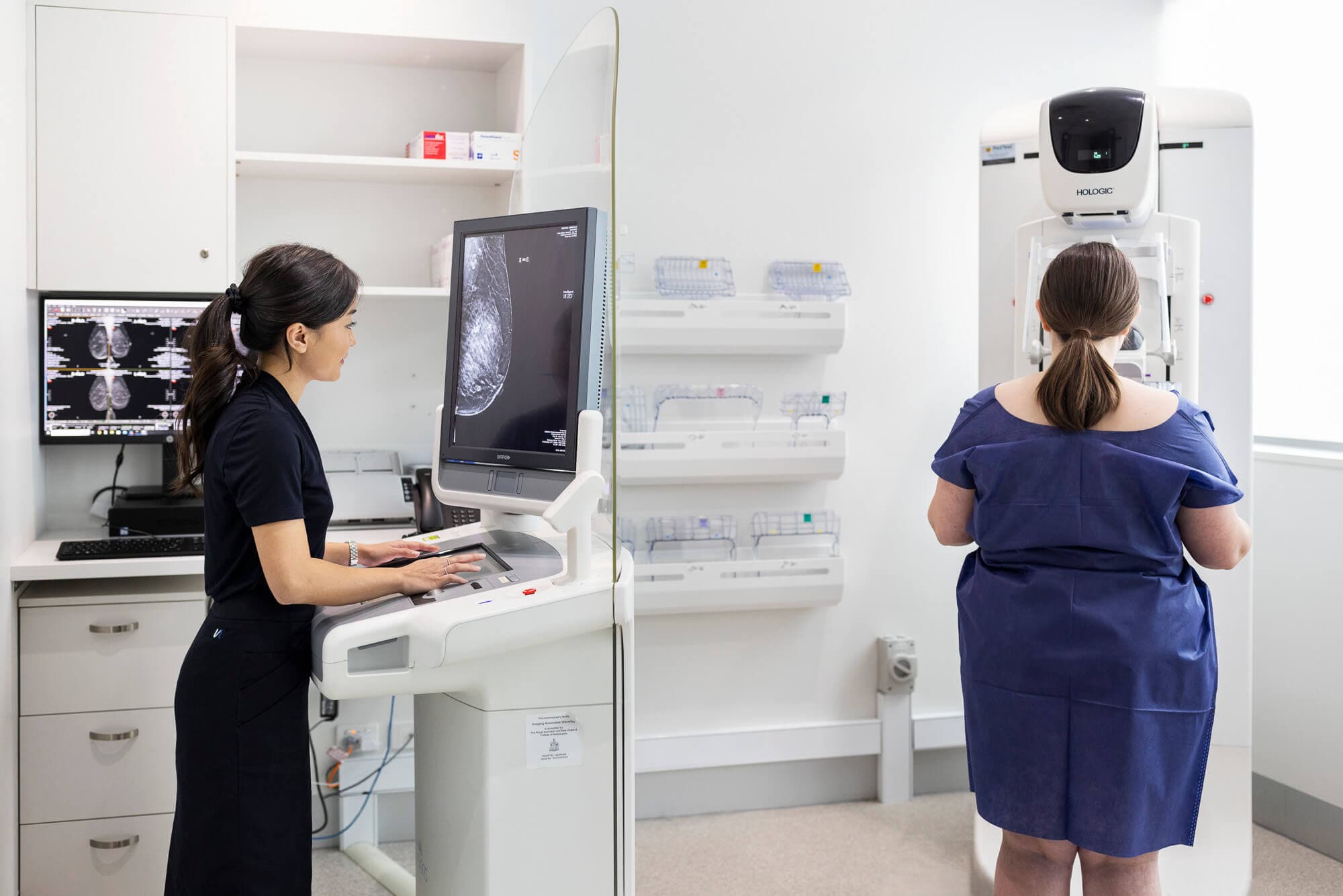
We are proud to offer a comprehensive range of both ultrasound and tomosynthesis guided breast biopsy procedures, including vacuum assisted breast biopsy (VABB), performed by our specialist breast radiologists.

Breast Biopsy Procedures:
Ultrasound Guided Breast Biopsy
Ultrasound imaging can be used by a radiologist to guide a needle to the correct location to take a tissue sample. A biopsy may be requested of an area of possible abnormal tissue which needs to be further characterised.
The Radiologist will perform this procedure. Sometimes there may be a
Sonographer and/or Nurse in the room at the time. The radiologist will clean the skin surface in the area that the biopsy will be made and you will be given an injection of local anaesthetic, which can cause a mild sting.
Once the area is numb, the Radiologist will use the ultrasound probe to
visualise the biopsy needle entering into the breast tissue. Once in position, the
Radiologist will use the biopsy device to remove small amounts of breast tissue. You will usually feel some pressure and hear a clicking noise as the biopsy is taken. This is then sent to a pathologist for analysis and diagnosis.
Generally there is no preparation for this Breast Biopsy procedure. Specific information will be given to you at the time of booking.
Please advise staff at time of booking if you are on any medications, particularly blood thinners.
Tomosynthesis Guided Breast Biopsy
Your mammogram may identify an area of breast tissue that requires further investigation. Using the mammogram x-rays, the radiologist can guide a biopsy needle into the exact location of the area of concern. A biopsy may also be performed under 3D Mammogram to further assist in the location of the area of concern.
You will be asked to change into a gown which allows your chest to be accessed
easily for the scan. You will be in a chair for the examination and your breast
will be compressed between two plates on a specialised x-ray unit. The
compression not only holds the breast still, but also thins out the tissue to make
the picture clearer. Low dose x-rays are used to produce an image.
During the procedure, the radiologist will clean the skin surface in the area that the biopsy will be made and you will be given an injection of local anaesthetic, which can cause a mild sting. These biopsies are often performed with vacuum assistance, and a clip marker is often placed in the breast at the site of biopsy at the end of the procedure.
There is no preparation required for this procedure. Please advise us of any
medication you are taking prior to your scan.
Breast Clip Marker Insertion
Sometimes a clip marker will need to be placed in your breast under ultrasound or tomosynthesis guidance following a biopsy. This is to ensure the relevant area can be found again if you are required to have surgery, in which case the clip marker is usually removed during the operation. If you do not need surgery, the clip marker remains in your breast.
The clip marker is tiny (about the size of a grain of rice), and you will not feel it in your breast. It is made from a surgical titanium alloy, similar to surgical clips commonly used in other operations.
How do I book a Breast Biopsy Procedure near me?
Schedule an Interventional Breast Procedure by completing our booking form or calling your nearest Imaging Associates radiology clinic now. Breast Imaging Services are available at the below Imaging Associates locations.
- Melbourne VIC – Mitcham, Box Hill
- Wagga Wagga NSW – Edward Street
Imaging Associates accepts all medical imaging referrals, even if written or printed on another radiology company’s request form.
Book now
In this section
CT ScanLung Cancer ScreeningAbdomen CT ScansCT Pulmonary AngiogramCT Coronary AngiogramCT Calcium ScoreCT Lumbar SpineDental X-RayX-RayDEXA ScanFluoroscopyInterventional Breast ProceduresInterventional Radiology: Sports, Spinal and Pain Management3D MammogramMagnetic Resonance Imaging (MRI)Nuclear MedicineUltrasoundPregnancy UltrasoundWomen’s Imaging
Book an appointment
Complete our booking form to request an appointment at an Imaging Associates radiology clinic near you.
Book now


